- By: CE4RT
- Patella PA
- Patella PA Oblique Lateral Rotation
- Patella PA Oblique Medial Rotation
- Patella PA Axial Oblique Kuchendorf Method
- 膝蓋骨接線投影 Hughston Method
- Patella Tangential Projection Merchant Method
- Patella Tangential Projection Settegast Method (Sunrise View)
- Patella Skyline Laurin View
- Suggested Reading
- RLKA
By: CE4RT

This article for the Radiologic Technologist (X-Ray Tech) discusses radiographic positioning of the knee for patella views.
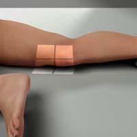
Patella PA
Purpose and Structures Shown To show the patella in PA projection. This view shows greater detail in the patella due to the decreased OID (object to image receptor distance).
Position of patient Prone position with the legs extended.
部分の位置 IRの中心を膝蓋骨に当てる。 膝蓋骨がIRの平面と平行になるように脚を調整します。
中心線 IRと膝窩部に対して垂直で、膝蓋骨から出る。
技術的要素 軟部組織の可視化、大腿骨遠位部の鋭いトラベキュラーマーク、および膝蓋骨の明確な輪郭は、動かずに最適な照射ができることを示します。 重なっている大腿骨を通して膝蓋骨を見るには、十分な貫通が必要です。
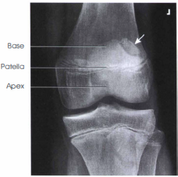
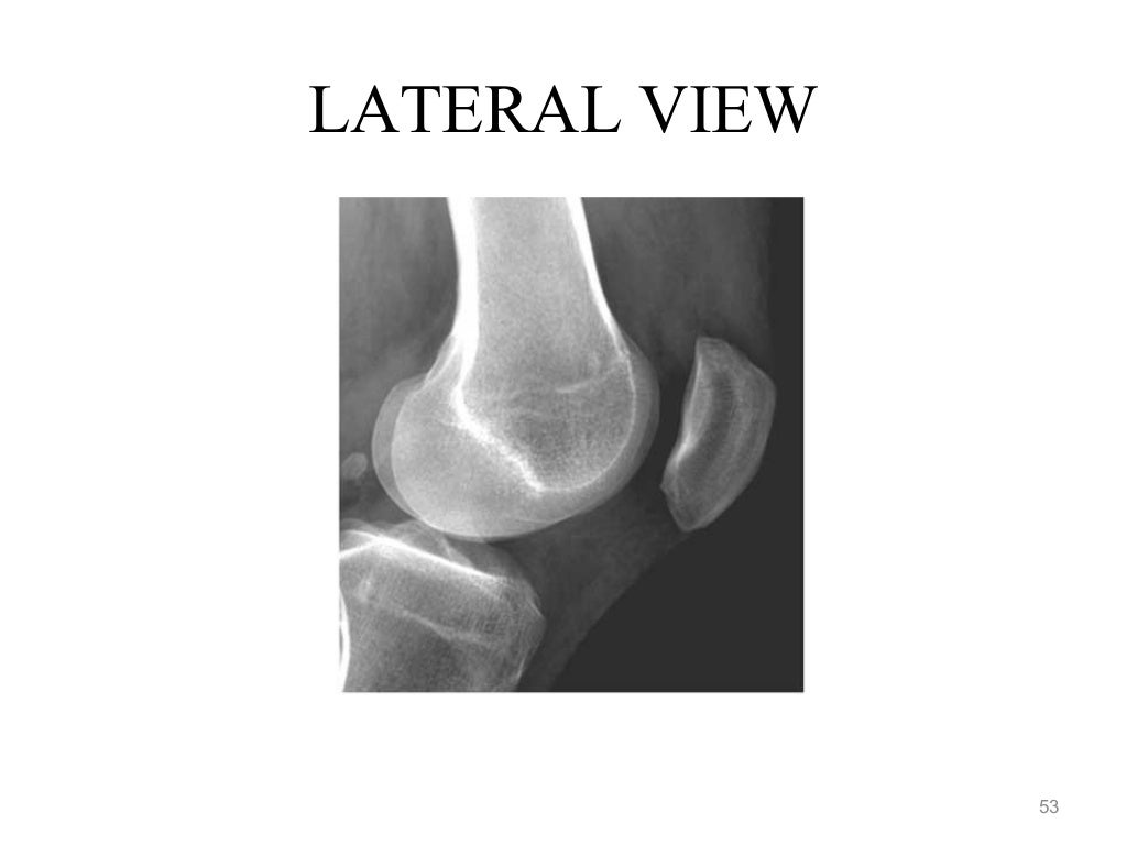
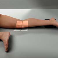
膝蓋骨の側面
目的と構造 膝蓋骨の側面プロファイルと膝蓋大腿関節腔を示すものです。
Position of patient Lateral recumbent position(患者の体位)…横向きになった状態。
部位位置 患者に患側の股関節を向くように指示します。 支持のために足首の下に砂袋が置かれるかもしれない。 影響を受けた膝を約5~10度曲げる。 屈曲を大きくすると、膝蓋大腿関節のスペースが狭くなる。
中心線 IRに垂直で、膝蓋骨から膝に入る。
インソール-サルバティ比 膝蓋骨の位置を評価するために用いられる比率である。 X線側面写真で、膝蓋骨の対角線上の最長距離と膝蓋腱の長さの比率である。 この比率は、膝関節の屈曲の程度に関係なく測定することができます。 A ratio of less than 0.8 is indicative of patella alta (high-riding patella).

Patella PA Oblique Lateral Rotation
Purpose and Structures Shown Medial margin of the patella superimposed over the femur and also the lateral aspect of patella free of the femur.

Position of patient Prone position.
Position of part Flex the knee 5-10 degrees. Externally (laterally) rotate the knee 45 -55 degrees from prone position.
Central ray Perpendicular to the IR, exiting the palpated patella. When the patella is being imaged, collimation should be to the mid-knee area.
Patella PA Oblique Medial Rotation
Purpose and Structures Shown Majority of the medial patella without the femur.

Position of patient Prone position.
Position of part Flex the patient’s knee about 5-10 degrees. Medially rotate the knee 45-55 degrees from the prone position.
Central ray Perpendicular to the IR, exiting the palpated patella. Collimate closely to the patellar area.
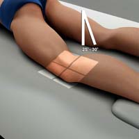
Patella PA Axial Oblique Kuchendorf Method
Purpose and Structures Shown Majority of the patella is seen free of superimposition by the femur.
Position of patient Prone position(患者の位置) 横臥位。 患側の股関節を2~3インチ高くする。 足首と足の下に砂袋を置く。 膝がわずかに曲がるように(約10度)調節し、筋肉をリラックスさせる。
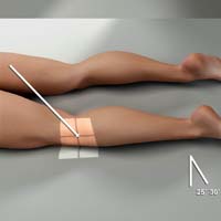
部分の位置 膝蓋骨へのIRを中心にする。 直立伏臥位よりも楽な伏臥位から膝を約35~40度側方回転させます。 人差し指を膝蓋骨の内側縁に当て、横方向に押します。 膝を前内側で休ませ、膝蓋骨を側方にずらした状態で保持する。
中心線は、膝蓋骨と大腿顆の間の関節空間に、尾側25~30度の角度で向け、膝蓋骨の後面に入る。
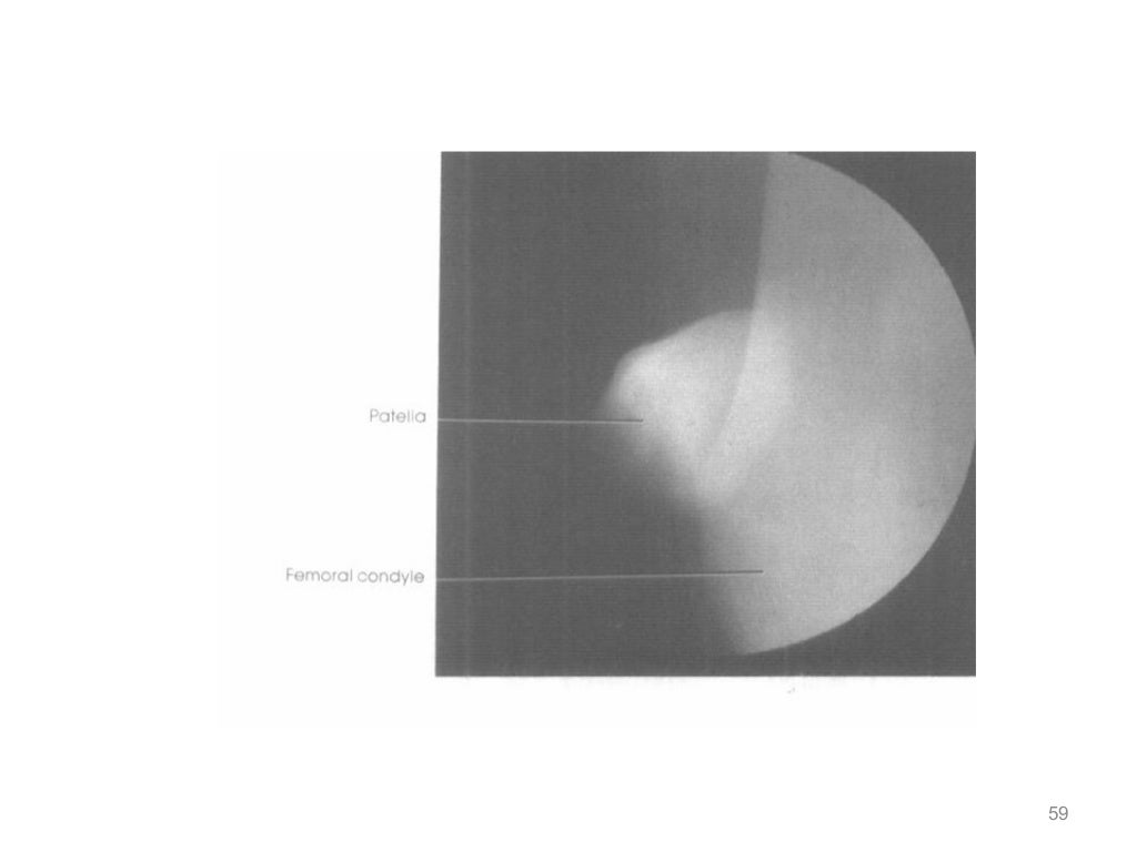
膝蓋骨接線投影 Hughston Method
目的および構造示す 大腿顆を挙げることができます。 この投影は、膝蓋骨の亜脱臼と膝蓋骨骨折を示します。
患者の位置 足をテーブルの上に置いて、うつ伏せの姿勢。
Position of part IRを患者の膝下に配置します。 脛骨と腓骨がテーブルから50~60度の角度を形成するように、患部の膝をゆっくり曲げる。
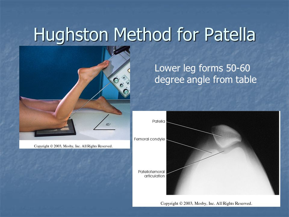
Central ray 頭側および下側に45度角度付け、膝蓋大腿関節を通して指向させる。
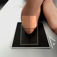
Patella Tangential Projection Merchant Method
目的と示す構造 膝蓋骨と膝蓋大腿関節を軸方向に投影し、わずかに拡大した画像として見ることができます。
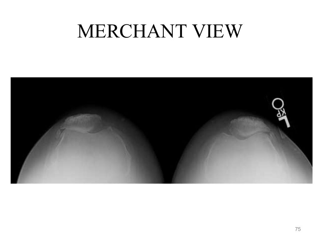
Position of patient supine with both knees at the end of table.
テーブルの端に両膝を置いて、仰臥位にします。 膝と下肢を調節可能な赤外線保持装置で支持する。

部品の位置 “axial viewer” 装置を使用して、患者の膝を約2インチ上げ、大腿骨がテーブルトップと平行になるようにします。 膝の屈曲角度を40~45度に調整する。 両足をふくらはぎの高さでひもで縛り、脚の回転をコントロールし、患者をリラックスさせる。 IRをCRに対して垂直に置き、膝蓋骨から約1フィート遠位で患者の脛骨に当てます。 患者がリラックスしていることを確認します。 大腿四頭筋の弛緩は、正確な診断のために重要です。 リラックスしていないと、亜脱臼した膝蓋骨が顆間溝に引き戻される可能性があり、その結果、正常と誤認されることになります。
中心光線 IRに垂直な光線。 ビームは尾側と下側に向けられる。 膝関節屈曲45°でCRを水平面から30°尾側(垂直から60°)に傾け、CRと大腿骨の角度を30°とする。 CRは膝蓋大腿関節の高さで膝蓋骨の中間に入ります。

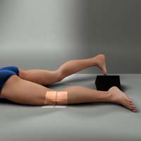
Patella Tangential Projection Settegast Method (Sunrise View)
目的と表示構造 膝蓋骨と膝蓋大腿関節に関する情報を提供します。 膝蓋骨のアキシャルイメージを得るための代替法である。 一般的な手法は、後方視投影法です。

Position of patient supine or prone (prone is preferred because the usually can be flexed to a degree and immobilization is easier)仰臥位またはうつ伏せ。 仰臥位ではIRを膝の下に置き、膝を水平軸から115度曲げます。

患者がX線撮影台に座っている場合、患者は膝の後ろでIRをしっかりと保持する必要があります。

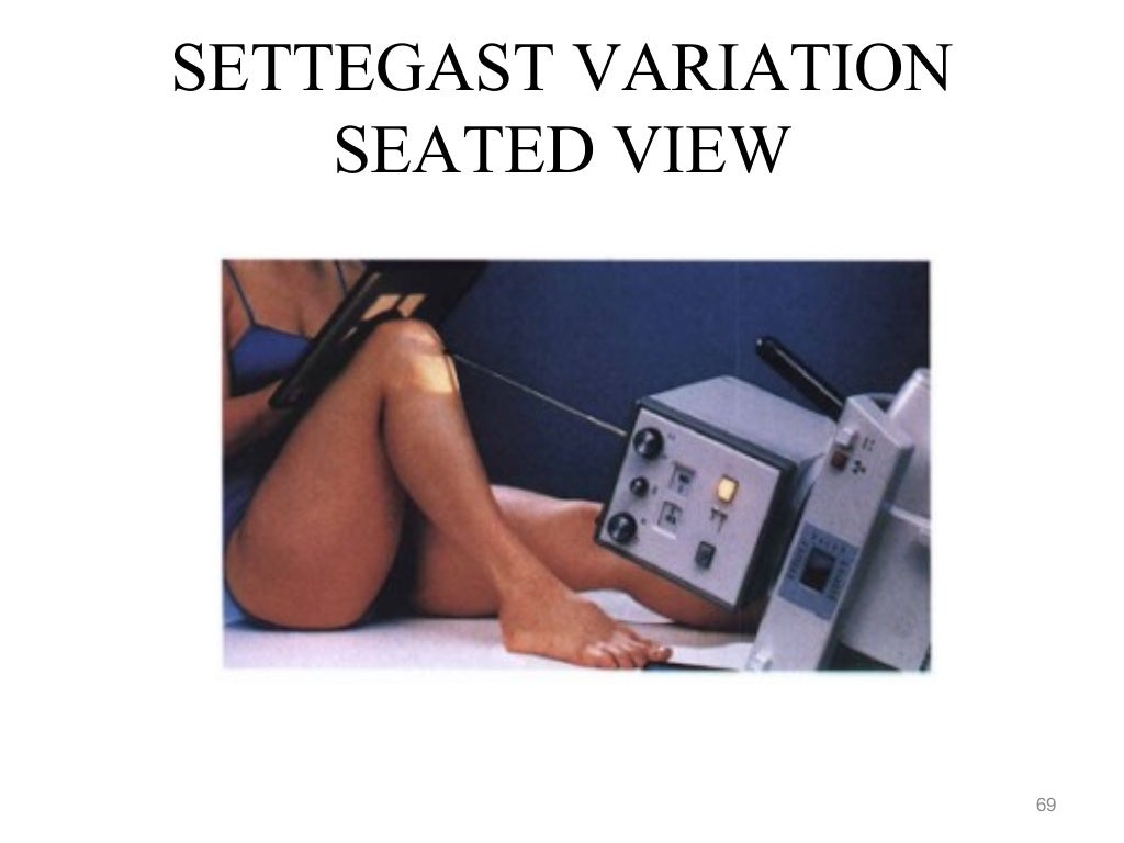
Position of part 患者の膝をできるだけゆっくり、あるいは膝蓋骨がIRに対して垂直になるまで曲げる(患者の状態が許す場合)。 ゆっくり均等に曲げれば、患者はその位置に耐えることができるが、速く不均等な曲げ方をすると過度の痛みを引き起こす可能性がある。 必要に応じて、長い包帯を患者の足首または足に巻きつけます。 患者に肩の上で紐の端を掴んでもらい、脚の位置を固定します。
中心線 関節が垂直のとき、膝蓋骨と大腿顆の間の関節腔に垂直であること。 そうでない場合は、膝の屈曲の度合いによってCRの角度の度合いが変わる。 通常、角度は下腿から15~20度程度になります。

Patella Skyline Laurin View
目的と示される構造 これは、骨構造による重畳を排除して可視化した膝蓋骨の下垂体軸投影である。 膝蓋大腿関節が明瞭に確認できる。 スカイライン投影では、他のビューでは見逃される可能性のある膝蓋骨の外側面の骨折が確認できる。

Position of patient semi-recumbent on the examination table with an pillow or cushions behind the back to enable the position to maintain the patient is the position. 患者は横向きで膝蓋骨の上のIRを保持する。

Position of part ひざは20~30度屈曲させています。

Central ray The beam is directed cephalad and superior, 160 degrees from the vertical axis or 30 degrees from the horizontal axis. The X-rays pass inferior to superior through the patella. The tube will need to be below the level of the table.
Suggested Reading
Radiography of the leg and knee: An overview of anatomy and pathology
Radiographic positioning of the leg
Radiographic positioning of the knee in AP views
Radiographic positioning of the knee in PA views
Radiographic positioning of the knee in lateral views
RLKA
Read more about this and other subjects and get 2 Category A ARRT CE Credits in the X-Ray CE Course “Radiography of the Leg, Knee, and Ankle”